Experiments
Biomechanical characterization of brain tissue
Biomechanical testing of ultrasoft tissues such as brain tissue is challenging as the response highly depends on length and time scales, the loading mode, strain and strain rate, or drainage conditions. Our goal is to establish an experimental procedure that minimizes falsifying effects, but also sufficiently covers loading scenarios relevant for real-life applications. We complement rheological, nanoindentation, and atomic force microscopy experiments performed at the LTM, FAU, with triaxial biomechanical testing at the Institute of Biomechanics, TU Graz, led by Prof. Gerhard A. Holzapfel, to thoroughly characterize animal and human brain tissue. Hand in hand with the mechanical modeling, we optimize the experimental procedures to assess the information required to establish realistic constitutive models and accurately calibrate the corresponding material parameters.
In addition to native brain tissue, we investigate biopolymeric substitute materials in collaboration with the Chair of Biomaterials, FAU, led by Prof. Aldo Boccaccini and the Biophysics group, FAU, led by Prof. Ben Fabry, see also [Link].
Analysis of the correlation between mechanics and microstructure
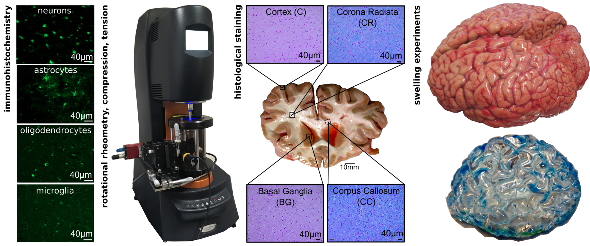
To obtain a basis for microstructurally motivated constitutive and evolution laws for brain tissue, we analyze the microstructure of mechanically tested specimens through histological staining, immunohistochemistry, and immunolabeling in collaboration with the Chair of Anatomy, FAU, led by Prof. Friedrich Paulsen. Our goal is to identify the microstructural components dominating the macroscopically recorded tissue response. A rheometer equipped with a microscope module allows us to track individual microstructural components such as cell nuclei during mechanical loading, as illustrated in Figure 1.
Analysis of the evolution of properties during development, homeostasis, and disease
In addition to the characterization of brain tissue behavior at a certain point in time, we aim to understand the evolution of properties during development, homeostasis, or disease. Through novel experimental procedures, we quantify how both mechanical properties and structure change with time – autonomously or induced by mechanical load. Swelling experiments as illustrated in Figure 1, for instance, allow us to reproduce the evolution of the macroscopic structure of our brain.